


Modern visual recovery operations are high-tech and safe procedures that enable us to eliminate almost all ophthalmic problems.They have been successfully used for decades, so these methods are constantly evolving, expanding and becoming more effective.Visual functions can be improved by hardware correction of the shape of the cornea, lens, retina and other elements of the eye optical system.The rightly chosen technology not only completely restores vision, but also reduces the risk of complications.From this article, you will learn about what ophthalmic procedures, instructions for use, and possible risks.
type
Vision and minimally invasive programs are reliable and have minimal invasiveness due to the development of medical hardware methods.They last no more than a few hours and there is no need to take complicated rehabilitation measures in the future.The choice of surgical treatment depends on the patient's visual device's disease, age and general condition.
Laser calibration
The most popular type of operation to correct vision.These are high-tech methods today with minimal complication risk.Allows you to deal with myopia, hyperopia and astigmatism.After the operation, maintain visual acuity for a long time, and if you follow all instructions from your ophthalmologist, you can avoid repeated interventions altogether.There are several types of laser correction:
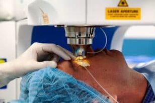
- Lasik.The basic type of operation for restoring vision.First, the surface layer of the cornea is separated by micromagnets and then changed using laser rays.The main disadvantage of this correction is that it is not possible to consider individual characteristics of the patient's eye anatomy.
- Super Lasik.Advanced version of traditional LASIK methodology.You can take into account the structure of the patient's visual system to enable you to achieve better results.Used in most modern clinics in the world;
- femto lasik.For similar types of operation, the only difference is that the cutting of the cornea is not performed by a micro movement, but by a special Femo laser.There is an improved version where the procedure of the operation depends on the individual characteristics of the patient - Super Femto Lasik;
- Epi-lasik.The mechanism of this procedure is the same as that of the traditional LASIK method, but this procedure is only directed to patients with corneal thinness (acquired or congenital).
- PRK (FRK).Photorefractive keraectomy has been performed since 1985.Today, in the presence of contraindications, it is suitable for common correction methods, such as having subtle corneal, severe ophthalmic diseases.The recovery process is always painful and the recovery period lasts longer than other methods.
The visual correction operation lasts no more than 15 minutes.After the operation, protective dressing must be worn for several hours and dripped for 1-2 months.The risk of complications is small, requiring repeated treatment and significantly reduced vision.
Vitrectomy
This is a procedure for removing the vitreous body of the eye.It is performed under general or local anesthesia and in the absence of complications it passes within 2-3 hours.First, a small puncture was performed in the eye socket, followed by subsequent operations.Usually, this is the recovery of the retina-affected area of laser, foreign densification or fabric integrity.This procedure is specified for the following issues:
- Recovery of visual function after eye tissue bleeding;
- Prevent age-related retinal detachment;
- Treatment of severe retinopathy that occurs with severe scarring or neovascularization (vascular germination).
Artificial polymers, bubbles, silicone oils or balanced salt solutions are used as alternatives to vitreous bodies.The latter type is often used because there is no need for repeated operations in the future - the salt solution is subsequently replaced with intraocular fluid.
After the surgery, side effects occur in the form of swelling of the corneal, increasing intraocular pressure and even more vision.Recovery and prediction depend on the widespread lesion and the type of prosthesis when replacing the vitreous body.If there is an irreversible change in the optic nerve, visual correction is almost impossible.
Sclerosis
A common ophthalmic surgery designed to strengthen the eye shell (sclera).Its regulations are not to correct visual function, but to stabilize the degree of myopia in the risk group.It is recommended that it be performed by adolescents with this problem, because at this age, the shape of the eye is changing positively.
During operation, the required material of the sclera is strengthened and the flaps are introduced behind the posterior wall of the eyeball.Polymers or biological ingredients are usually used.After that, spikes in the outer shell of the eye, and after a few months, the blood vessels needed to maintain visual function grow into the skin flap.There is a simplified curing membrane shaping procedure.It is an introduction to artificial or biological matter of the eye.The mechanism of action in this technique is the same - preventing the growth of the eyeball.
Sclerosis
This is a carefully studied operation that will not actually change over the years.It is done in most clinics.There are actually no side effects except allergies to medications.A second operation is usually required.
Replace lens
The necessary operation is specified in any other degradation process in the cloud or in the lens.Always forced treatment, but the implant is selected individually, depending on age, gender, and the severity of pathological changes in the eye.Crystalus substitution is specified in the following cases:
- The height of myopia and hyperopia;
- Refractive index is significantly reduced;
- Regeneration process in the eyes, vision-related reduction;
- The impossibility of laser vision recovery;
- cataract;
- Possibility of developing glaucoma in the context of systemic or ophthalmic diseases.
This process is always carried out under local anesthesia.During the procedure, the surgeon uses a laser to make a small incision, after which the patient's lens is diluted with a special tool and removed from the eye.Afterwards, the prepared transplant was installed.The intervention lasts no more than 25 minutes, followed by no need for seams and recovery in the hospital.
The surgery is performed in most private and state clinics.There are usually no complications after manipulation, but subsequent laser vision correction is usually prescribed.In rare cases, the lens needs to be replaced again.
Keratogenesis (replacement of corneal)
One of the most modern and complex ophthalmic procedures, associated with many risks, requires the high quality of the surgeon.The anatomical integrity and physiological function of the cornea are needed to be restored.It is a provision for the treatment of congenital or acquired defects obtained by injury or disease.Healthy transplant fabrics are only performed in donors, but are developed with artificial alternatives in many countries.Keratopathy is recommended to solve the following problems:
- Treatment of corneal disease (Belko, tone disorder);
- Mechanical or chemical damage;
- Congenital defects.
The operation shall not exceed 30 minutes.During the procedure, a surgeon with a laser or special scalpel can remove a portion of the patient's cornea and implant the donor fabric into its location.The seams can last until one year, after which special lenses are selected to reduce the risk of infection.The recovery period starts at 4 weeks, during which antibiotics are required, but the subsequent year requires regular checkups.
In recent years, due to the use of special ingredients during processing and storage, the risk of donor fabric rejection is likely to be greatly reduced.
Retina laser coagulation
How to restore retinal tissue.The method is more than 70% effective and can already return to the usual lifestyle 24 hours after implementation.Within one year after the operation, observations must be performed in the observation of an ophthalmologist.
Today, the operation is done with a laser, which allows you to do it without bleeding.Anesthesia is performed locally and the process time is not more than 20 minutes.
Before exposure to the laser, the drip drips to expand the pupil and then a special protective lens is placed through which it is exposed at low frequencies.Due to the high temperature, the bonds are damaged cells as well as small blood vessels.
The coagulation process is necessary for any damage and pathology of the retina, as well as the eyes and diseases of the vascular system of the organ.After surgery, inflammation and shadowing may occur.In the years after the correction, it is impossible to engage in a lot of physical labor and active exercise.
Cross-linking
An effective method for treating various corneal diseases.It is intended to enhance ligaments and other fibers in corneal tissue, which is necessary for malnutrition for keratoconus of varying degrees or degenerate processes.
The procedure was performed under local anesthesia.First, a small portion of the cornea is cut off with a special device and vitamin B2 is infused into the open area.The subsequent radiation allows you to compact the fabric over 200%.In the first week after the operation, it is necessary to wear protective contact lenses and be examined by a doctor for 6 months.The effect of this process lasts for 10 years and then requires repeated operations.
Complications are observed in rare cases.Patients may reduce vision, inflammation, or shadows in the cornea.
Glaucoma treatment
When drug therapy does not bring the necessary results, various ophthalmic procedures for glaucoma are required.This procedure is performed using laser or surgery.
The laser method is considered to be the most successful.He is completely painless to the patient and has almost no complications.During the process, a hole is made with a beam to remove liquid from the tissues of the eye to normalize the pressure.It is used to treat all types of glaucoma.
The manual surgery type is less efficient because there is a risk of complications after surgery.Usually, this is a non-erupt deep sclerosis resection.The purpose of this method is to use a slightly sparse corneal layer to reduce intraocular pressure.
As time goes by, the effect will decrease after both types of operations.On average, after 5-7 years, a second operation is required.Qualified medications can be used to extend this period.
in conclusion
Today, in most modern ophthalmic clinics, many surgical procedures are performed for vision correction.These are accurate high-tech methods that you can solve for almost any eye defect.The choice of this method depends on many factors - age, disease, individual characteristics of the patient's visual system structure.After the surgery, the effect occurs almost immediately, and if prescriptions from all doctors are observed, your vision can be preserved for a long time.